Case Study: Congenital Peritoneopericardial Diaphragmatic Hernia

A 16-week-old entire male, Springer Spaniel puppy was presented to our out of hours provider having collapsed. The owner reported that earlier that day, he vomited up his breakfast, was shaking and lethargic, by the evening he was weak and could not get up.
Clinical examination
The patient was weak, tachycardic (160bpm) with hypodynamic pulses, tachypnoeic (60bpm) and hypothermic (35.6°C). His mucus membranes were pale pink with a prolonged capillary refill time of 3 seconds.
Initial treatment
A shock treatment plan was provided with aggressive fluid therapy and intravenous antibiotics.
Diagnostics
Blood tests showed a stress leukogram and hyperlactatemia (11.1mmol/L). POCUS ultrasound revealed no free fluid in abdomen, but the presence of intestines in the chest cavity was detected. Radiography performed once transferred back to Portland (primary care vets) showed presence of abdominal organs within the thorax, confirming ultrasonographic findings.
Diagnosis
Peritoneal-Pericardial Diaphragmatic Hernia (PPDH) is a congenital condition, where there is remaining communication between the pericardial and peritoneal spaces through the diaphragm. The patient was also found to have an umbilical hernia and only one testicle was descended in scrotum, both conditions commonly seen with PPDH. The treatment of this condition is indicated when the patient becomes symptomatic, with signs usually referring to herniated abdominal contents. The prognosis is good following surgical correction. Referral for urgent specialist surgical correction was recommended but declined due to prohibitive cost. As the alternative was euthanasia, in-house surgery was offered at Portland Vets. Risks included anaesthetic-related complications and surgical complications such as haemorrhage, tissue breakdown, surgical complexity, which is unable to be corrected, pneumonia, pleural effusion and inability to self-ventilate following intra-operative intermittent positive pressure ventilation (IPPV).
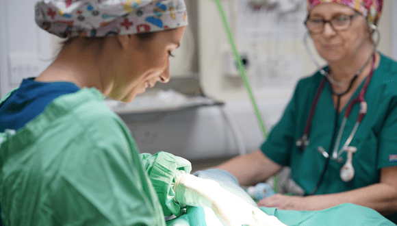
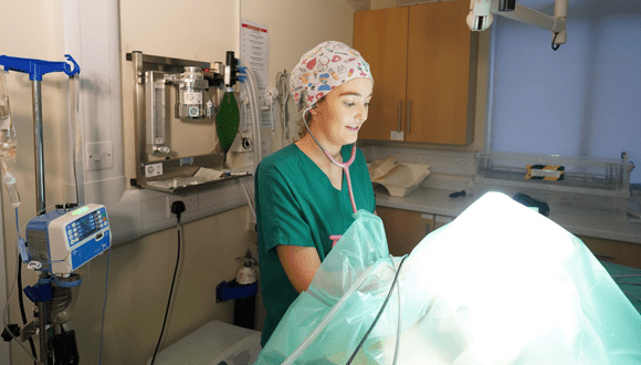
Treatment
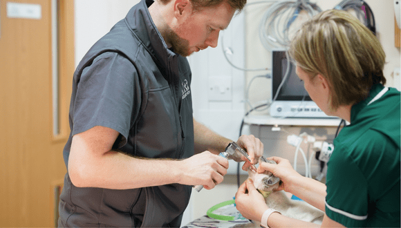
A mildly elevated pain score indicated the need for buprenorphine administration. Intravenous cephalosporin antibiosis and maropitant were also administered. The dog was induced with IV alfaxalone following premedication with midazolam and methadone. The patient was maintained with isoflurane and oxygen therapy and a bupivacaine splash block in the incision was performed. A midline incision was made and a 5-10cm diaphragmatic defect was found with a portion of small intestines and omentum herniated and showing first signs of strangulation.
A part of strangulated omentum was removed, and the portion of intestines was confirmed to be viable after replacing them in the abdominal cavity.
A small amount of free fluid was suctioned out of pericardium and the heart was inspected. The pericardium was incised to allow drainage and aid with diaphragmatic defect closure, which was closed with non-absorbable suture, the last of the air was suctioned out using a urinary catheter restoring negative intra-thoracic pressure The abdomen was closed routinely, repairing the umbilical hernia at the time.
Outcome
The patient was recovered uneventfully with normal respiratory rate and effort throughout. A chest drain was judged to be unnecessary due to normal ventilation peri and post-operatively as well as having the potential to become a complication due to interference from the young puppy. The patient maintained normal vital parameters throughout the recovery and in the 24 hours post-surgery, he started eating and regained activity within hours. Parenteral maropitant and opioid analgesia was maintained after the surgery and a 5-day course of oral cephalosporin and metronidazole was started. The patient was discharged the following day and re-presented for post-operative checks at days three and ten.